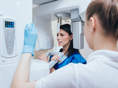
Digital radiography refers to the use of electronic sensors and computer systems to capture, display, and store dental radiographic images. Unlike traditional film, digital systems convert X-ray energy directly into a digital file that can be viewed immediately on a monitor. This technological shift has changed how clinicians evaluate oral health, allowing for more informed, timely decisions during an appointment.
For patients, the most noticeable difference is the speed and clarity of the images. A digital radiograph appears on screen seconds after exposure, enabling the dental team to review findings in real time and explain them visually. The immediacy of this process improves communication and helps patients better understand diagnoses such as cavities, bone loss, or developmental concerns.
From a clinical standpoint, digital radiography is a diagnostic tool that supports precision. Clinicians can zoom, adjust contrast, measure structures, and compare images on the spot — actions that are difficult or impossible with conventional film without additional processing. These capabilities make radiographs more useful for treatment planning, monitoring healing, and documenting long-term changes.
Digital sensors come in a few formats, including intraoral sensors that sit inside the mouth and extraoral panels used for panoramic or cone beam imaging. Intraoral sensors are thin and designed for patient comfort while producing high-resolution images of individual teeth and surrounding bone. The sensor’s sensitivity and pixel density determine how much detail the image contains, and advances in sensor design have markedly improved image quality over the last decade.
Once an image is acquired, software tools help clinicians enhance diagnostic value without re-exposing the patient. Adjustments such as changing brightness and contrast, applying filters, or magnifying a region of interest can reveal subtle findings that might be missed on a raw film. Because these manipulations are non-destructive to the original file, records remain intact for future reference or comparison.
Integration with the electronic patient record is another advantage. Images are stored directly in the patient’s chart, tagged with dates and notes, and organized for easy retrieval. That organized workflow reduces the risk of misplaced films and ensures that diagnostic images are always available when needed for follow-up care or referrals.
One of the primary benefits of digital radiography is reduced radiation exposure compared with conventional film. Digital sensors require less radiation to produce a clear image, and modern X-ray units are calibrated to use the lowest dose necessary for diagnostic quality. This dose reduction is especially valuable for children and patients who require periodic imaging over time.
Beyond lower exposure per image, digital systems support conservative radiographic practices. Because clinicians can view and evaluate images instantly, repeat exposures due to under- or overexposure are less common. In addition, targeted imaging techniques — taking only the views that answer a specific clinical question — help limit cumulative dose without compromising care.
Safety also extends to image handling and storage. Eliminating film eliminates the need for chemical developers, which reduces environmental hazards associated with processing solutions. Digital archives replace physical film storage, decreasing clutter and the potential for film degradation over time.
Digital radiography enhances clinical diagnostics by making it easier to detect caries, assess periodontal bone levels, evaluate root anatomy, and locate impacted teeth. Because the images can be manipulated and measured precisely, clinicians can make more accurate assessments and plan treatments with greater confidence. This precision is useful across general dentistry, restorative procedures, endodontics, and oral surgery.
Another practical advantage is improved collaboration. Digital images can be shared quickly with specialists, laboratories, or insurance providers as needed for consultations or to coordinate care. When a referral is required, transmitting digital files avoids delays and ensures the receiving clinician has the necessary visual information to proceed efficiently.
Documentation is also strengthened. Consistent, time-stamped images provide an objective record of a patient’s oral health over time. This historical perspective helps clinicians track disease progression or healing after treatment and supports clinical decisions with clear visual evidence.
For most patients the experience of having a digital radiograph taken is straightforward and comfortable. The sensor is positioned briefly inside the mouth or the machine is aligned externally for panoramic views. Because images display immediately, the appointment does not require the wait associated with film processing, and the dentist can review results with the patient during the same visit.
Clinicians will explain the reason for each image and how it contributes to diagnosis or treatment planning. Visual aids on the monitor make it easier to demonstrate issues and outline next steps in language that is easy to follow. This transparency supports shared decision-making and helps patients feel informed about their care options.
If follow-up images are needed, clinicians will use the minimum number necessary and will select imaging parameters appropriate for the patient’s age and clinical condition. Patients with particular concerns about radiation should feel empowered to discuss them; the dental team can outline the diagnostic value of the proposed images and the measures taken to keep exposure as low as reasonably achievable.
In summary, digital radiography represents a practical, patient-centered evolution of dental imaging. It delivers clearer diagnostic information faster, reduces environmental impact, and supports safer, more collaborative care. If you'd like to learn more about how digital imaging is used in our office or have questions about what to expect during your next visit, please contact us for more information.
Digital radiography uses electronic sensors and computer processing to capture X-ray images and convert them directly into digital files. Instead of exposing film, a sensor records X-ray energy and the image appears on a monitor within seconds. This immediacy allows clinicians to review and discuss findings with patients during the same visit.
Captured images can be enhanced, magnified, and measured using software tools without altering the original file, which preserves diagnostic integrity. The digital format also facilitates organized storage and easy retrieval in a patient’s electronic health record. Together, these features streamline diagnosis and clinical decision-making.
Unlike conventional film, digital radiography eliminates chemical processing and produces images almost instantly, reducing both wait times and environmental hazards from developer solutions. Digital sensors typically require less radiation exposure to generate a diagnostic-quality image, and software tools make it possible to enhance image detail electronically. These differences improve workflow efficiency and expand the range of diagnostic information available during an appointment.
Another contrast is in record keeping and sharing: digital files are stored electronically and can be backed up, tagged, and retrieved far more reliably than physical film. Clinicians can compare current and past images side by side on the screen, which supports monitoring of disease progression or healing. Overall, the transition to digital improves convenience, safety, and clinical utility.
Digital radiography is considered safe when used appropriately because modern sensors require lower doses of radiation than conventional film to produce clear images. X-ray units used with digital sensors are calibrated to deliver the minimum exposure necessary for diagnostic quality, and clinicians follow principles that limit unnecessary imaging. This dose reduction is particularly important for children and for patients who need periodic follow-up images.
In addition to using low-dose equipment, dental teams employ targeted imaging techniques to capture only the views needed to answer a specific clinical question. Protective measures such as lead aprons or thyroid collars are available when clinically indicated and help further reduce exposure. Patients with concerns about radiation are encouraged to discuss them with the dental team so imaging decisions can be individualized.
Dental practices commonly use intraoral sensors for bitewing and periapical images, and extraoral panels or cone beam systems for panoramic and three-dimensional views. Intraoral sensors are thin and shaped to fit comfortably inside the mouth while capturing high-resolution images of individual teeth and surrounding bone. Extraoral systems provide broader anatomic context useful for orthodontic assessment, implant planning, and surgical evaluation.
Sensor technology varies in sensitivity and pixel density, which influences the level of detail in the image, and manufacturers continue to improve sensor design for comfort and diagnostic performance. Software compatibility and image formats are also important considerations, as they affect integration with patient records and the ability to share images with specialists. Clinicians select the sensor type based on the diagnostic need and the patient’s comfort.
Digital radiographs help clinicians detect cavities, assess periodontal bone levels, evaluate root morphology, and locate impacted or missing teeth, among other diagnostic tasks. Software tools allow precise measurements, contrast adjustments, and magnification to reveal subtle findings that might be missed on raw film. These capabilities increase diagnostic confidence and allow for more targeted treatment planning.
Because images are available immediately, dentists can correlate radiographic findings with the clinical exam in real time and explain concerns visually to patients. Time-stamped digital records also support tracking changes over time, which is useful for monitoring disease progression or healing after treatment. This combination of visual evidence and documentation strengthens clinical recommendations.
Digital images are typically stored within a patient’s electronic health record and are tagged with dates and notes for easy retrieval. Practices use secure server systems and backup protocols to protect patient records from loss, and access controls limit who can view or modify images. These measures reduce the risk of misplaced films and ensure continuity of care across visits.
When images are transmitted for referrals or laboratory work, they are usually sent using encrypted channels or secure specialist portals to maintain patient privacy. Retention policies follow regulatory guidelines so that records remain available for clinical follow-up while preserving confidentiality. Patients may request copies of their images or ask that files be forwarded to another provider as needed.
The imaging process is generally quick and comfortable: intraoral sensors are positioned briefly inside the mouth for periapical or bitewing views, while the machine is aligned around the head for panoramic images. Because the image appears on-screen almost immediately, there is no waiting for film processing and the dentist can review results with the patient during the same visit. Staff will explain the purpose of each image and how it contributes to diagnosis or treatment planning.
If additional images are needed, the clinician will minimize the number and adjust exposure settings appropriate for the patient’s age and condition. Patients who have concerns about radiation or the procedure are encouraged to raise them; the dental team will explain the diagnostic benefit and the safety measures in place. Clear communication helps patients understand what to expect and makes shared decision-making easier.
Yes. One of the practical advantages of digital images is the ability to transmit them quickly and accurately to specialists, laboratories, or referring providers. Electronic files can be sent via secure channels, attached to referral packages, or uploaded to specialist portals so the receiving clinician has immediate access to the necessary visual information. This streamlined sharing reduces delays and supports coordinated care.
When collaborating on a case, clinicians can annotate images, include measurements, and provide clinical notes that clarify the reason for referral. Digital sharing also avoids the need to rely on physical film, which can be lost, degraded, or require additional processing. Maintaining clear documentation and secure transmission preserves patient privacy while enabling efficient consultations.
High-resolution digital images allow for precise assessment of tooth structure, root anatomy, and bone levels, all of which are critical for planning restorative, endodontic, or surgical procedures. Measurements and image manipulation enable clinicians to select appropriate techniques, anticipate challenges, and design customized treatment approaches. These tools support more predictable outcomes and reduce the likelihood of unexpected complications.
Digital records also facilitate long-term monitoring by providing consistent, time-stamped images that track healing or disease progression. When combined with other digital workflows—such as digital impressions and CAD/CAM restoration design—the imaging data helps create integrated treatment plans that are efficient and well-documented. Overall, digital radiography contributes to a higher standard of evidence-based care.
Maplewood Dental Group integrates digital radiography into routine exams and treatment planning to ensure timely, evidence-based decisions for patients. Imaging is used selectively to answer specific clinical questions, whether detecting decay, evaluating periodontal health, or planning restorative and surgical care. The practice stores images in the electronic chart so findings are accessible for follow-up visits and collaborative consultations.
During appointments clinicians review images with patients on-screen to clarify diagnoses and outline treatment options in straightforward terms. If a referral or laboratory input is needed, digital files are transmitted securely to support efficient coordination of care. Patients are encouraged to ask questions about any proposed imaging so that decisions reflect both clinical need and personal preferences.
Skilled, compassionate dental care for patients of all ages in Nashua, New Hampshire.